
Clinical standard MRI images obtained using Dedicated 3T Extremity Scanner
The initial integration of our next generation superconducting magnet, together with our gradient coils and Radio Frequency (RF) coils was completed in late 2017 and images obtained. In collaboration with our system integration partner, work to increase the level of integration between the system elements and to enhance the system capabilities has continued. These activities have resulted in the acquisition of clinical-quality images for knee, wrist and ankles from a dedicated extremity 3T (Tesla) imaging system based around a magnet utilising Magnetica’s proprietary IP. This outcome represents a significant milestone in Magnetica’s commercialisation journey.
A dedicated extremity MRI scanner provides cost savings for the operator during both installation and throughout its operational lifetime whilst increasing patient comfort.
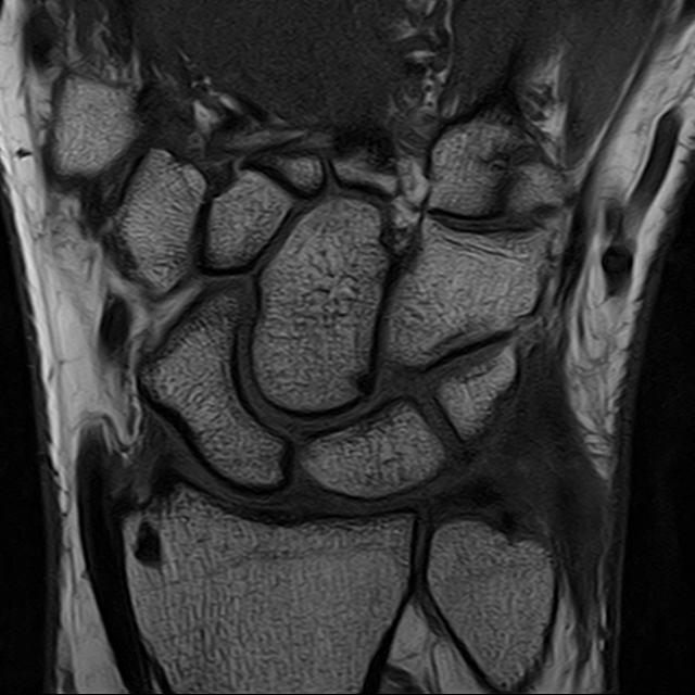
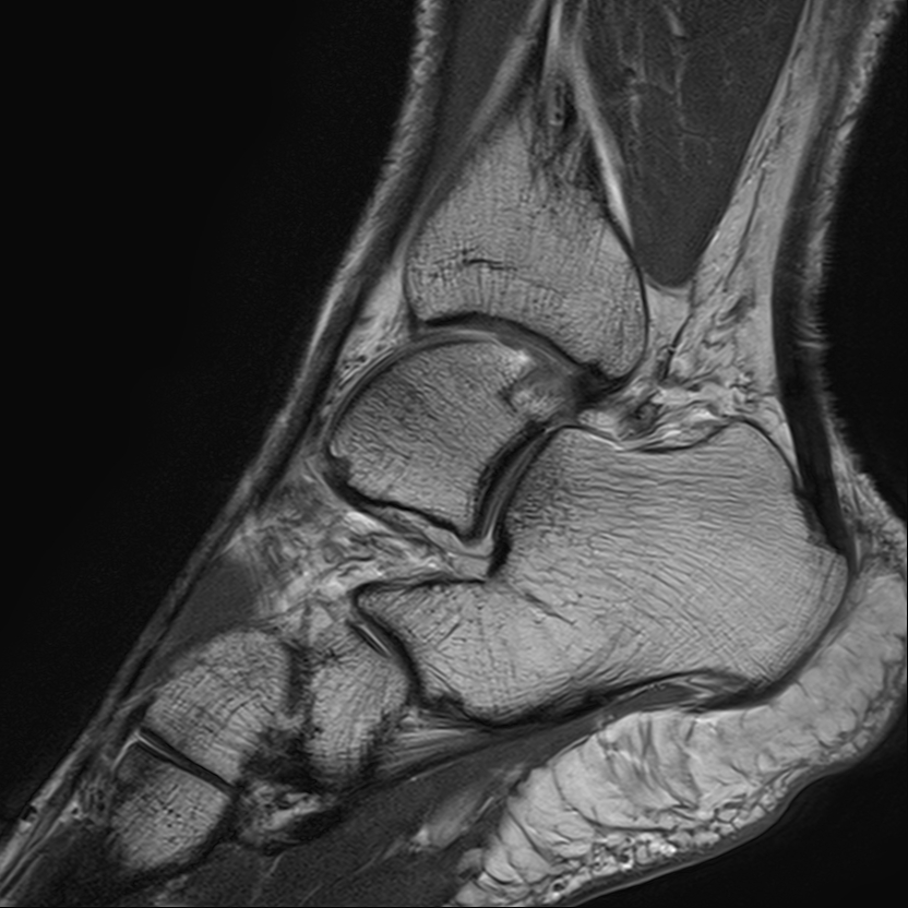
Images: Wrist and Ankle scanned using 3T dedicated Extremity MRI system